
 Camden

 Contact us for
 Contact us for
Hair Loss
Hair Loss
Hair loss can have many different causes. Excessive loss is when more than 100 strands of hair is lost a day. Hair loss can occur in patches (localised), from all over the scalp (diffuse) or in pattern.
Androgenetic alopecia (patterned hair loss) is the most common hair loss condition and can occur in both men and women. The tendency to develop androgenetic alopecia runs in families and is almost inevitable in men.
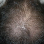
To slow down the hair loss, both men and women can consider the following options
Topical therapy
Finasteride for men, and a few types of oral medications for women
Low level laser light therapy
Hair Transplant surgery
Alopecia areata (localised hair loss) most often results in circular, coin-sized patches on the scalp. Oftentimes, the hair regrows by itself. It can also affect the entire scalp with dramatic hair fall which is extremely distressing for the patient. Your doctor may perform blood tests to check for any underlying conditions. Treatment involves topical medication, injections, light treatment and oral tablets if necessary.

Telogen effluvium (diffuse hair loss) commonly occurs after severe stress to the body. Physical stress results after childbirth, high fever and severe infections, inadequate diet and dramatic weight loss, major surgery, and underlying illnesses. Emotional stress can also trigger telogen effluvium as well certain oral medications. Your doctor will take a detailed history and perform blood tests if needed. Fortunately, the hair loss stops on its own often within a few months. Medications can help correct any contributing conditions and shorten the period of hair loss.
While awaiting recovery or regrowth of hair, numerous natural options are available to allow temporary camouflage of your hair loss condition so that you may not feel embarrassed in public.
There are many other types of hair loss conditions and scalp disorders. Please see your dermatologist for an accurate diagnosis and appropriate treatment.
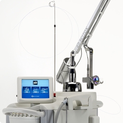
Laser Hair Removal (Long Pulsed Nd:Yag laser and IPL)
Effective hair removal is done via the following systems, Cutera Excel HR, XEO IPL, Sciton ClearScan Long Pulsed Nd:Yag laser, Sciton BBL (IPL). Depending on the skin and hair type, and the location of the hairs, a series of treatment sessions are recommended to achieve the desired result.
How does laser hair removal work?
The laser used in hair removal targets the hair follicle, thus damaging the follicle and removing the hair with minimal damage to the surrounding skin.
What areas of the body can be treated?
Our laser hair removal is safe in all skin types, enough to be used on all body parts including the face, upper lip, back, underarms, arms, bikini line and legs.
What can I expect with the procedure?
Laser hair removal gives a prickly or a light rubber-band snapping sensation. Usually, a topical anesthetic cream is applied 30 to 45 minutes before treatment. No special precautions are needed after the treatment. Some of the treated hairs would be burned off immediately although most of the treated hair will not fall out until 2 weeks later. Mild redness and swelling after the treatment is expected. These reactions subside within few hours to 1 – 2 days.
How long does each treatment take?
An upper lip and chin can be done in 5 minutes. Underarms or bikini lines usually take 10-15 minutes. Legs and back take longer.
Are there any possible side effects?
Side effects are few and rarely permanent. Skin lightening (hypopigmentation) is rare, but darkening (hyperpigmentation) may occur, especially in patients with darker skin tones. These skin colour changes will fade with time.
Is the hair removal permanent?
Permanent reduction in hair growth is achieved in 70 – 80% of individuals treated, and the best results are achieved after multiple treatments. You may have a few hairs re-grow over a period of time so touch-up treatment may be needed for removal from time to time.
How many treatments do I need?
Hair goes through different growth phases and lasers work most effectively against hairs that are in the active growth phase. Initially, it is recommended that you undergo a series of four to eight treatments at four to eight-week intervals depending on the area treated.
The ClearScan Long Pulsed Nd:Yag laser is also useful for eliminating the appearance of redness and fine vessels on the skin and also helps with non-invasive rejuvenation of the face
Please visit www.sciton.com/products/clearscan-yag and www.cutera.com for more information.
Skin Infections
Molluscum Contagiosum
Molluscum contagiosum is an infectious viral disease of the skin caused by the poxvirus. Infection is common in children, especially those with eczema. Transmission can occur via direct skin or mucous membrane contact.
Molluscum are smooth pink, or flesh-colored, dome-shaped, umbilicated papules. Most patients have many in common areas such as the underarm, popliteal fossae, and groin. They often self resolve but occasionally can persist and spread.
Treatment options include cryosurgery, laser ablation, curettage, cantharidin, topical creams.

Viral Warts
Warts are common and are due to the human papillomavirus (HPV).
The common wart is the most frequently seen as a hyperkeratotic, flesh-colored papule or plaque studded with small black dots. Other types of warts include flat warts and plantar warts.
Treatment options include cryosurgery, electrosurgery, laser ablation and topical lotions. Sexual partners of patients with genital warts will require examination with possible need for the HPV vaccines. These vaccines ( Gardasil) aim to prevent further genital HPV infection and ultimately cervical cancer in women.

Herpes Simplex
Herpes simplex virus (HSV) infection is a painful, and often recurrent blistering infection. Common areas include the lips, the mucosa of the mouth and genital areas. Sometimes your doctor may take some of the blister fluid for testing.
Acyclovir and valacyclovir are effective. For recurrent infection (more than six episodes per year), suppressive treatment is warranted.
Herpes Zoster ( Shingles)
Shingles is an acute, painful blistering rash that affects up to 20% of adults. It begins with pain in a bandlike area on the skin, followed by redness and blisters a few days later.
It is due to the reactivation of the varicella virus and often occurs after illness, stress, trauma, and surgery.
Treatment includes rest, painkillers and early antiviral therapy. There is a vaccine ( Zostavax) that is available to prevent the occurrence and severity of zoster outbreaks.
Tinea infections (Fungal)
Fungal infections of the skin are common in our hot and humid climate. It can affect any part of the body from the toenails to the scalp. They exist in various forms and often appear as flaky scaly patches which may be red or itchy
Tinea pedis (athlete’s foot) is the most common fungal infection. Tinea cruris (jock itch) occurs in the groin and on the upper, inner thighs and buttocks. Tinea capitis, or fungal infection of the scalp, is most common in children. Tinea corporis (body), faciei (face), and manuum (hands) represent infections of different sites of the body. Tinea unguium (onychomycosis) is fungal nail disease, characterized by thickened yellow nails and subungual debris.
Tinea (Pityriasis) Versicolor
Usual areas include the back, chest, and neck. The rash appears and circular scaly pale or brown patches on the skin which may be itchy.
Treatment options include antifungal creams, shampoos and washes. If needed, oral anti-fungal tablets are recommended.
Bacterial infections
Impetigo is a skin infection usually caused by Staphylococcus aureus and occasionally by Streptococcus pyogenes. It appears a weepy areas of the skin, drying to a yellowish pink crust with occasional blisters.
Folliculitis is a infection of the hair follicles appearing as red painful or itchy firm bumps or pustules.
Erysipelas and cellulitis appear as red warm painful swollen areas of the skin, often on the fingers, hands, feet or legs, and requires early medical attent
Mole Removal and Minor Skin Surgery
Moles, also known as beauty spots or birth spots, may be present at birth or more commonly arise later on in life. They may be flat, raised or even be hair-bearing. Most moles that have not changed in terms of their characteristics are harmless. Occasionally, a mole has to be removed because there is a risk it has become malignant (cancerous) or because it causes some discomfort (for example catching on clothes or a razor).
Various methods are used to remove moles. They include surgical removal by excision or using laser removal. Surgical excision is recommended in slightly larger and more protruding moles. Stitches may or may not be necessary depending on the size and location of the mole.The recovery period from surgical removal is about a week. Patients can usually carry out their normal activities during this period. Laser removal is recommended for flatter moles. A carbon dioxide laser &/or a pigment laser is used for this procedure. Recovery of the laser wound takes about 5 days and normal daily activities may be carried out during this period.
Your dermatologist will advise on the method of mole removal is most appropriate for you.
Skin Cancers
Skin cancers are one of the top 10 most common cancers in Singapore (Singapore Cancer Registry 2004-2008). It can be caused by many factors including excessive sunlight exposure and a decrease in a person’s immune system. In recent times, there has been a rising trend in the incidence of skin cancers, likely from an increase in sun-seeking behaviour or activities, coupled with increasing life expectancies in our population. Often times, potential skin cancers may present early in its precancerous or in-situ form. An example of this is Actinic Keratosis.
Some common types of skin cancers include:
Basal Cell Carcinoma
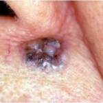
This is the commonest form of skin cancer in human beings. It presents as a slow growing growth or ulcer on the skin. The aid of a dermatoscope may be helpful in diagnosing this condition in suspicious cases.
Squamous Cell Carcinoma
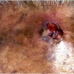
This presents as a firm irregular fleshy growth usually on sun exposed skin or on the legs. The growth can increase rapidly in size giving rise to a large lump which may sometimes break down to form an ulcer. If untreated, the cancer may spread to the surrounding lymph nodes.
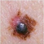
Malignant Melanoma
This is a cancer arising from the melanocytes (pigment cells) of the skin. It is one of the most aggressive and potentially life-threatening skin cancers. It presents as a discoloured or pigmented spot that may display one or more of the following features:
Becomes Asymmetrical in shape
Its Borders become irregular
It displays more than 2 Colours within a single growth
There’s Discomfort (or soreness) on the spot
The spot rapidly Enlarges
Melanomas can occur on any part of the skin
Treatment For Skin Cancers
All skin cancers should be removed soonest. Precancerous lesions such as Actinic Keratosis can be treated with liquid nitrogen or creams. The confirmatory diagnostic test for skin cancers is a biopsy.
For established skin cancers, complete surgical excision is the treatment of choice. Clearance Margins on Histology with surgery ensures very high cure rates for most skin cancers.
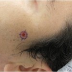

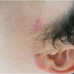
Occasionally, non-surgical treatment options with special creams, cryotherapy or curettage may be considered. In cases of malignant melanoma, special blood tests, scans or other tests may be required for staging & treatment of the condition.

Pigment Laser (Q-switched Nd:YAG laser)
We use the MedLite® C6 laser and the Spectra XT laser (Q-switched Nd:YAG laser) to target the excess pigment in the skin. Light is absorbed by the unwanted pigment target. The pigments then break up and disappear.
What can I expect from the procedure?
An anesthetic cream is usually applied on the area to be treated 30 mins before treatment. The procedure itself is rather painless. Occasionally, some patients experience a mild pricking sensation or feel some warmth over the treated area. Depending on the type of pigmentation treated, one may experience some redness or mild crusting after the procedure which can last 3 to 5 days
How many treatment sessions will it take to remove the pigmentation or tattoos?
Depending on the type of pigment in your skin, it can be gone in as few as one to two sessions, especially with freckles and sun spots (lentigenes). Certain pigmentation such as Hori’s nevus or certain birthmarks may require repeated treatment sessions. Similarly with tattoos, the average tattoo requires anywhere between 4-8 treatments. Treatment sessions are usually spaced 6 – 8 weeks apart.
Are there any side effects?
The treated area may darken sometimes but will fade within 2-3 months. This is part of the healing process and lightening cream may be prescribed for you to speed up recovery. Other side effects such as skin lightening and scarring are fortunately rare.
Common Benign Skin Growths
There are many types of benign skin growths. Most can be treated quite easily, or possibly even left alone. Below are some common skin growths.
a.Keratosis (Seborrhoeic)
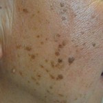
These are brown to black growths on the face or body. They may increase in number or size. Sometimes, it mimics a skin cancer. They can be removed quite easily with minor surgery or by laser.
b.Skin Tags
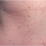
These are skin coloured lumps that grow out of the skin. They are soft and usually found on the neck, armpits and body. These are harmless but can be removed by minor surgery or by laser.
c.Milia Seeds
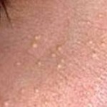
These are oil ‘seeds’ that are trapped just under the skin. They are found predominantly on the face. They can be removed by simple expression in the clinic.
d.Sebaceous Hyperplasia
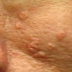
They are enlarged oil glands on the face which appear as small yellow irregular lumps. They are found predominantly on the face. They can be removed by minor surgery or by laser.
e.Syringomas
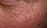
These appear as small skin-coloured bumps usually on the eyelids and cheeks. They are benign growths of the sweat ducts that are painless. They can be removed by minor surgery or by laser.
f.Viral Warts


These are common harmless skin growths caused by a virus. They can occur on any part of the skin, both in the young & old. There are various treatment options. One option is liquid nitrogen therapy. Repeated treatments may be required to eradicate the wart completely. Alternatively, they can be removed by minor surgery or by laser.

Picosecond Laser
The Candela Picoway® delivers high peak power within the shortest pulse durations using photoacoustic laser energy rather than photothermal laser energy to fracture pigment particles. This is done with minimal damage to the surrounding skin tissue. This also allows lower energy and lesser number of treatments. Frequency of treatments range from once a month to once every 2 to 3 months depending on the condition being treated.
Indications:
Tattoo removal
Scars
Enlarged pores
Melasma
Lentigenes
Freckles
Rejuvenation
Please visit https://candelamedical.com/na/patient/product/picoway for more information
Diseases of the Nail: What Your Nails Can Tell About Your Health
A change in colour, texture, or shape of your nails can be harmless, but it can also be a sign of disease. Being aware of the health of your nails could give you an early warning of a serious problem that needs your attention.
Dark stripes down the nail
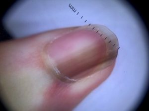
This is known as longitudinal melanonychia and is fairly common in people with darker skin.
If a fingernail or toenail has a new or changing dark streak, it’s time to see a dermatologist. That dark streak could be melanoma, the most serious type of skin cancer. Not every dark streak is a melanoma, but it’s always good to have a dermatologist examine one.
Oncholysis
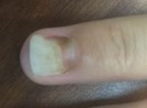
A common condition where the nail separates from the nail bed. This usually presents as a white discolouration at the tip of the nail.
The most common cause is trauma, including minor, repetitive trauma such as frequent tapping of long fingernails on a keyboard. However, fungal infections and psoriasis are also frequent causes.
Beau’s lines
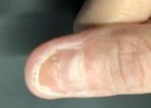
These are grooves or indentations that run across the nails. They can appear when the growth at the base of the nail is interrupted by injury or a severe illness. A fever, injury, chemotherapy, or major stress can cause your nails to grow slowly or stop growing. These grooves tend to be visible a few months after these conditions happen. That is when the nails have grown and the grooves have moved up the nails to become visible.
Thin, spoon-shaped nails (Koilonychia)
This occurs whenthe fingernails are thin and dip down in the middle and look like spoons. This could be caused by iron-deficiency anaemia.
Curved nails (Clubbing)

This occurs when the tips of the fingers enlarge and the nails curve around the fingertips. As the nails curve downward, fingertips often swell and the nails start to feel spongy when pressed on.
Curved nails can be a harmless trait, which runs in the family. Curved nails can also be a result of low oxygen levels in the blood, and could point to problems with the lungs, bowels or heart
Nail colour change
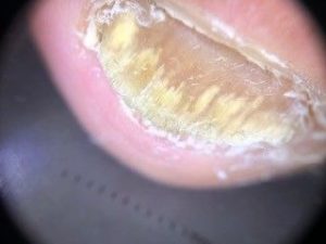
A change in the colour of the nails can portend adisease inside your body.
| Colour | Disease or other health problem |
|---|---|
| Blue nails | Not enough oxygen in your bloodstream |
| White nails | Liver disease, diabetes |
| Pale nails | Anaemia |
| Half pink, half white nails | Kidney disease |
| Yellow nailss | Lung disease, nail fungal infection |
| Dusky red half-moons | Can be caused by lupus erythematosus, heart disease, alopecia areata, arthritis, dermatomyositis |